Stages of Melanoma
Reviewed by: HU Medical Review Board | Last reviewed: May 2017. | Last updated: February 2023
Cancer staging is a way of describing how extensive a cancer is. The stage is related to tumor size. It also relates to whether the cancer has spread from the original tumor to other parts of the body. The cancer stage helps you and your doctor to plan treatment. It also provides information about survival.
There are many different ways of describing stages of cancer. The variations in method of staging have to do with differences between clinical guidelines and the associated research protocols that are part of how treatments are assessed. These differences are less important to you as a patient and more important to your healthcare provider in determining treatments or for tracking progress in a research study.
Melanoma is staged using a system that describes characteristics of the primary tumor, and the extent to which the cancer may have spread, either to lymph nodes, or other parts of your body. This staging system is referred to as the TNM staging system.1 This cancer staging system is used by most hospitals and medical systems.2 Once your doctor has categorized the T, N, and M, these values are combined to assign a cancer stage.
TNM Staging System for Melanoma
T categories
The T categories described below are ways of classifying the main tumor based on its thickness, ulceration, and mitotic rate.3 Your doctor gathers this information by doing a skin biopsy.
Thickness. The thickness of the tumor is described using the “Breslow Depth of Invasion” measure. Breslow thickness is a measure of how deep the lesion is from the top to the deepest point.4 It is measured in millimeters (mm). The number after the T (which ranges from 1 to 4) indicates the category of thickness.
Ulceration. Ulceration happens when the top layer of cells breaks down. This causes a hole to form in the skin, and the tissue below to show through.2 Ulceration indicates a worsening of the skin.5 The letter “a” at the end of the classification indicates no ulceration; the letter “b” indicates ulceration.
Mitotic rate. Mitosis is the process of cell division. The mitotic rate is the number of cancer cells that are in the process of dividing per square mm of the tumor area.6 Higher rates indicate a more problematic situation.6 Mitotic rate is used to classify very thin melanomas (T1; Breslow thickness less than or equal to 1.0 mm). The letter “a” is used when the rate is less than 1 per mm of tumor area, and the letter “b” is used when the rate is greater than or equal to 1 per mm of tumor area.2
Approximately 40% of melanomas are categorized as “Tis,” which is melanoma in situ (in place).7 At this stage, the cancer cells are only in the epidermis, the very top layer of skin. The tumor has not invaded deeper levels yet. At this stage, it is most common to treat and remove the melanoma by wide excision.7
N categories
The N categories indicate whether cancer cells have spread to the lymph nodes.3 N categories can be “clinically staged,” which means staged before lymph node surgery and range from 1 to 3. These stages are based on the physical exam and imaging tests.3
There are additional categories that can be used after surgery, when your doctor has a tissue sample. This is called “pathologic staging.” The pathologic stage indicates whether the melanoma is in the lymph node but so small it can only been seen under the microscope. This is called “micrometastasis” and is labeled with an “a.” The opposite is melanoma in the lymph node that is big enough to be seen on imaging or felt during the physical exam. This is called “macrometastasis” and labeled with a “b.” The letter “c” is used to indicate that the cancer cells have spread to nearby skin (satellites) or lymph vessels (in transit), but have not reached the lymph nodes.
M categories
The M categories indicate whether the cancer has spread to distant parts of the body.3 Simply, 0 means no metastasis and 1 means metastasis. Letters are added at the end to indicate the location of the metastasis.
The letter “a” is used to designate if the cancer has spread to other parts of the skin, tissue below the skin (also called subcutaneous tissue), or to distant lymph nodes. The letter “b” is used if the cancer has spread to the lungs, and the letter “c” is used if the cancer has spread to any other organ.
The M1c category can also be assigned when cancer has spread to any site of the body and when there is an elevated level of an enzyme called lactate dehydrogenase (LDH) detected in your blood. High LDH indicates cancer that is harder to treat.5
Putting it all together: assigning a stage
Once the cancer is characterized, the categories are combined to assign a stage.3 Generally speaking, the lower the stage, the better the chance for long-term cure and survival.
Pathologic Staging
Other classification and staging systems
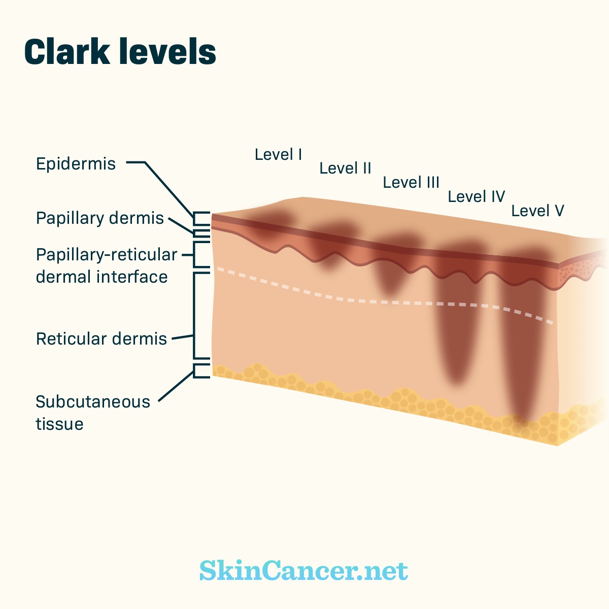
Melanoma is sometimes described and classified using other rating systems. One of these systems is called the Clark level. The Clark level is based on which layer of skin the tumor has invaded from the top layer of the skin (Level I) to invasion of the tissue under the skin, or subcutaneous tissue (Level V).4,8
Cancer registries may use more general categories to classify melanomas than those used in clinical staging systems. The terms in the table below are also useful in understanding cancer staging.4