Lymph Node Mapping and Biopsy
Reviewed by: HU Medical Review Board | Last reviewed: May 2017. | Last updated: April 2019
Lymph node biopsy is a procedure done to see whether skin cancer has spread. Tissue from the lymph nodes is studied under a microscope to see whether it contains cancer cells. Lymph node biopsy can be done in different ways.
How does melanoma spread?
Melanoma cells spread via the lymphatic system. Lymph is a fluid that contains white blood cells.1 Lymph travels through the body via lymph vessels.2 Lymph nodes are clusters of cells that filter the lymph, removing bacteria and other harmful substances.
Once melanoma has grown into the dermis layer of skin, it can enter the lymph vessels.3 If the cancer has started to spread, the closest lymph node is the most likely place to find it. Another term for the spread of melanoma to the lymph nodes is “regional metastasis.”
Figure 1. Lymph nodes and vessels

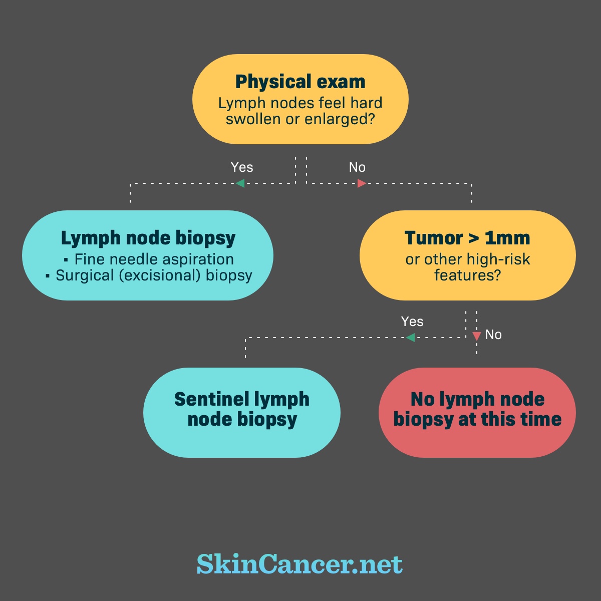
When is a lymph node biopsy needed?
Swollen, hard, and enlarged lymph nodes may be a sign that the cancer has reached the lymph nodes.4 Your doctor will feel your lymph nodes during the physical examination. Swollen lymph nodes may be described as “clinically positive,” “clinically detected,” or “clinically suspicious.” Biopsy is needed to confirm the clinical findings. Biopsy on clinically suspicious lymph nodes can be done in one of two ways:
- Fine needle aspiration biopsy
- Surgical (excisional) lymph node biopsy
The absence of lymph node symptoms does not rule out the spread of cancer. It is possible that the metastasis is so small that it can only been seen under the microscope. The term for this is micrometastasis.5 Based on the thickness of your tumor, your doctor might recommend a sentinel lymph node biopsy.4,6
Possible reasons for lymph node biopsy
Types of Lymph Node Biopsy
How is a fine needle aspiration biopsy done?
Your doctor will use a thin needle to remove tissue from your lymph node.3,4 If the suspicious lymph node is just under the skin, your doctor will be able find it by feel. If the lymph node is deeper, your doctor can use ultrasound or computed tomography (CT) scan to guide the needle. The area is numbed before the needle is injected. The procedure causes little pain and no scarring.3,4
Fine needle aspiration is accurate for finding melanoma. One estimate is that this procedure correctly identifies 92% to 96% of lymph nodes with melanoma.7 It also correctly rules out melanoma in about 99% of cases.7
How is a surgical (excisional) lymph node biopsy done?
Surgical lymph node biopsy is when the swollen lymph node or nodes are removed entirely. This procedure is also called excisional lymph node biopsy or open lymph node biopsy.4,8 Your doctor might recommend surgical lymph node biopsy if fine needle aspiration is not possible. It might be done if metastasis is likely, but the fine needle aspiration biopsy did not show cancer.
Surgical lymph node biopsy is performed in a hospital or outpatient surgical center.8 You may be sedated and given a local anesthetic (numbing medicine). General anesthesia might also be used, which means you will be unconscious for the procedure. Your surgeon will make a small cut and remove the swollen lymph node or nodes. Your surgeon will close the incision with stitches. It will leave a scar. You will probably be able to go home the same day. The lymph node goes to the lab to be studied.
How is sentinel lymph node mapping and biopsy done?
Sentinel lymph node biopsy is done to find microscopic cancer cells in a lymph node.3 You cannot tell by feel if there is microscopic cancer in the lymph nodes. Microscopic cancer also is too small to see with imaging tests.
Thick melanoma tumors are more likely than thin tumors to metastasize. Therefore, your doctor might recommend sentinel lymph node biopsy if your tumor is more than 1 millimeter thick.6 Sentinel lymph node biopsy is not routinely done when the melanoma is less than 1 millimeter.6 In such cases, the chance that the cancer has spread is very small.3
“Mapping” is done to determine how lymph travels around the tumor. The purpose is to identify the lymph node closest to the skin tumor, called the sentinel node. A radioactive dye is injected in the skin near the original tumor.4 A special camera is used to see where the dye travels. The place where the dye collects is the sentinel lymph node. Next, a blue dye is injected to make the vessels and sentinel lymph nodes stand out during surgery.9 A surgeon removes the sentinel lymph node and studies it under a microscope.4 If there are no cancer cells, then no more surgery is needed. If there are cancer cells, the rest of the lymph nodes in the area are removed. They are studied under a microscope. If cancer is found in the lymph nodes, more tests will be done to see whether it also has spread to distant organs.
How are the results of lymph node biopsy used?
The results of the biopsy are used to stage the cancer. Cancer staging is a way of describing how extensive a cancer is. The stage is determined by tumor size and cancer spread. Knowing the cancer stage helps with treatment planning. It also relates to your prognosis, or chance of survival.
The results might indicate that additional tests are needed. If the cancer is in the lymph nodes, it may also have spread to distant parts of the body. Your doctor may recommend imaging tests to look for cancer in the lungs, liver, brain, or bones.