What Does Basal Cell Carcinoma Look Like?
Reviewed by: HU Medical Review Board | Last reviewed: May 2017. | Last updated: February 2023
Basal cell carcinoma (BCC) develops when basal cells begin to grow out of control. Basal cells are found in the top layer of skin, called the epidermis.1 BCC grows slowly, and it rarely spreads to distant parts of the body. However, it must be treated. Untreated BCC can grow into bone or the tissue beneath the skin.1
BCC appears in many different ways. Descriptions of common presentations are below. It can be hard to identify a lesion correctly on your own. If you notice changes in your skin, discuss them with your primary care provider or dermatologist.
Where does basal cell carcinoma develop?
Skin that gets the most exposure to sun is most likely to develop BCC. BCCs are commonly found on the head, neck, and nose.1,2 However, about one-third of BCC tumors grow in areas that get little or no sun exposure.2 Rare cases of BCC develop around the anus.
Pictures of basal cell carcinoma
BCC has many different appearances. The three main forms of BCC are:3
- Nodular
- Superficial
- Morpheaform
These subtypes are based on how tissue samples look under the microscope. The subtypes have different appearances to the unaided eye, too. Up to 40% of tumors are a mix of two or more subtypes.2 Furthermore, some BCC tumors have melanin. Melanin is the pigment that gives skin its tan or brown color. Dark (pigmented) BCC is more common in people with dark skin tones than white people.4
Nodular BCC
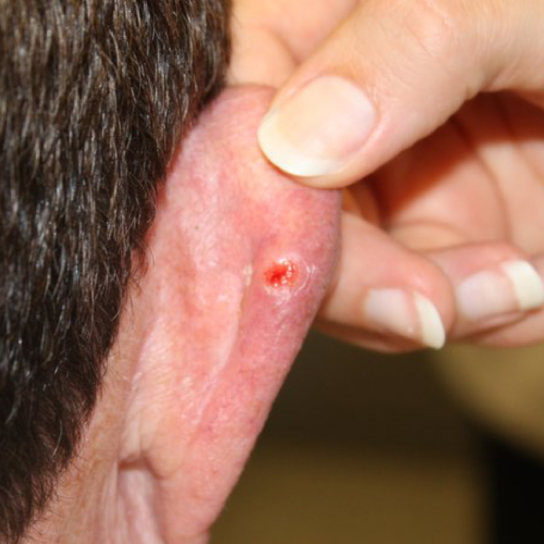
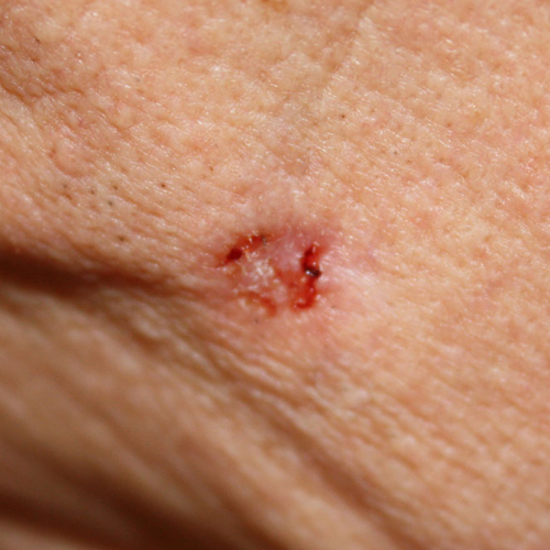
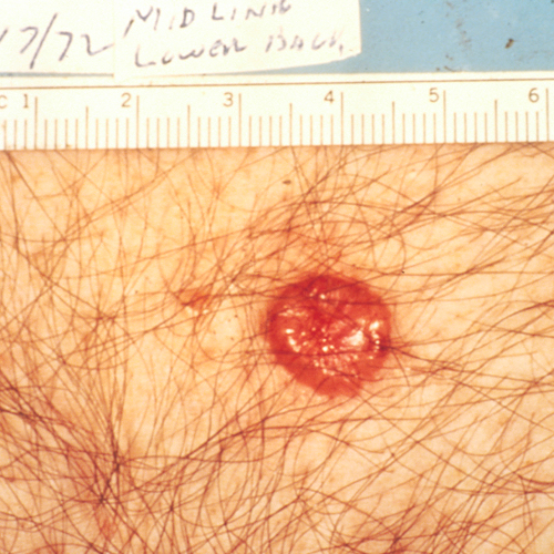
Nodular BCC looks like a dome-shaped bump. It may be pearly or shiny. Typical colors are pink, red, brown, or black. You may see tiny blood vessels in the lesion.2,3 These blood vessels cause the lesion to bleed easily. In fact, BCC is often mistaken for a shaving cut that will not heal.1
Over time, the top layer skin may begin to break down. This is called ulceration. Ulceration causes the center of the lesion to collapse. When this happens, the tumor looks like a crater.
Superficial BCC
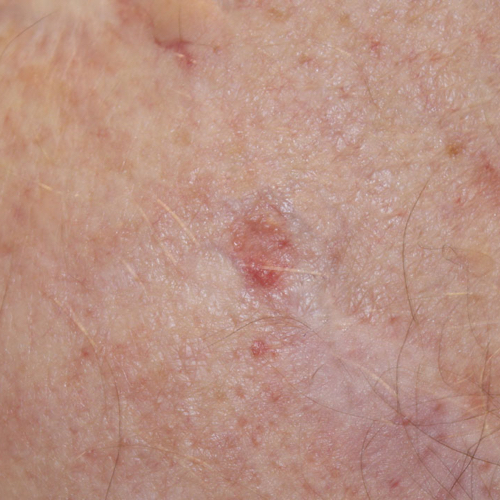
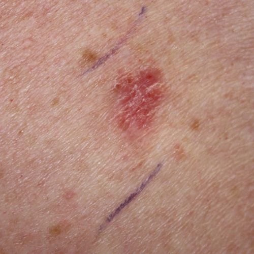
Superficial BCC looks like a scaly pink or red plaque.2,3 You may see a raised, pearly white border. The lesion may ooze or become crusty.3 Superficial BCC is typically found on the chest, back, arms, and legs.2
Morpheaform BCC
Morphea is a skin disease characterized by patches of hard skin. Morpheaform BCC got its name because it looks like morphea plaques.3 These waxy, light-colored lesions are hard, shiny, and smooth.2,3 They may look like scars. It can be hard to tell where the borders of these lesions are. Morpheaform BCC is often hard to spot.3
What are symptoms of basal cell carcinoma?
A lesion might be the only sign of BCC; you may not have any symptoms. The lesion may bleed easily. You might think it is a cut or sore that will not heal.
What else could this skin lesion be?
Other skin conditions may look like BCC. Nodular BCC without ulceration may look similar to:3
- Molluscum contagiosum, a viral infection that causes numerous small bumps.
- Sebaceous hyperplasia, a condition characterized by small yellow bumps.
- Intradermal melanocytic nevus, a nest of melanocytes in the dermis layer of skin.
- Fibrous papule, a firm bump that may develop on the nose.
- Other skin cancers (amelanotic melanoma, Merkel cell carcinoma)
Ulcerated BCC may be confused with squamous cell carcinoma or keratoacantoma.
Conditions that look similar to superficial BCC include:3
- Psoriasis
- Some forms of dermatitis
- Lichenoid keratosis, a condition characterized by small, inflamed patches or dark plaques
- Other skin cancers (amelanotic melanoma, Bowen’s disease) and precancers (actinic keratosis)
Morpheaform BCC may look similar to:3
- Scars
- Morphea (also called localized scleroderma)
- Other skin cancers (Merkel cell carcinoma, amelanotic melanoma, cutaneous adnexal tumors)
